Gram Positive And Gram Negative Bacteria : Schematic views of Gram negative (top) and Gram positive ... / Hence, appear in purple or hence, gram negative bacteria appear in the secondary stain colour which is pink.
Gram Positive And Gram Negative Bacteria : Schematic views of Gram negative (top) and Gram positive ... / Hence, appear in purple or hence, gram negative bacteria appear in the secondary stain colour which is pink.. Generally thinner, 11 to 15 nm. When comparing with the gram positive bacteria, gram negative bacteria are. Retain color of primary satin(crystal violet dye) and appears purple or blue. It paves way to the differentiation of the two distinct bacterial species. Gram positive bacteria retain the crystal violet stain during gram staining, giving the positive result.
Stabilised by teichoic acid and lipoteichoic acid. When the negative gram bacteria is stained with safranin or fuchsin in the experiment, it gives red or pink color. Gram staining technique is the most along with their staining characteristics, gram positive and gram negative bacteria differ from each other in various aspects which are listed below Gram positive bacteria vs gram negative bacteria (similarities and differences between gram positive and gram negative bacteria). Not sure if you need to know more for the scope of.
Understanding antibiotic resistance: 4.1 Gram-positive and ... from www.open.edu It accounts 50% or more of the dry weight of the wall of some gram positive bacteria. Diaminopamelic acid of one glycan backbone. Retain color of primary satin(crystal violet dye) and appears purple or blue. When comparing with the gram positive bacteria, gram negative bacteria are. The gram method allowed to divide all microorganisms into two large groups. Not sure if you need to know more for the scope of. They do not form a dispute, and in many cases are conditionally pathogenic. Gram positive bacteria vs gram negative bacteria (similarities and differences between gram positive and gram negative bacteria).
They retain the color of crystal violet and stain dark blue or purple.
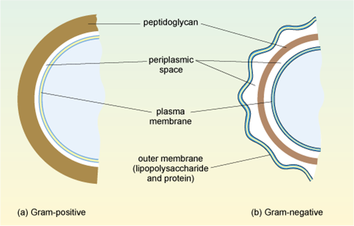
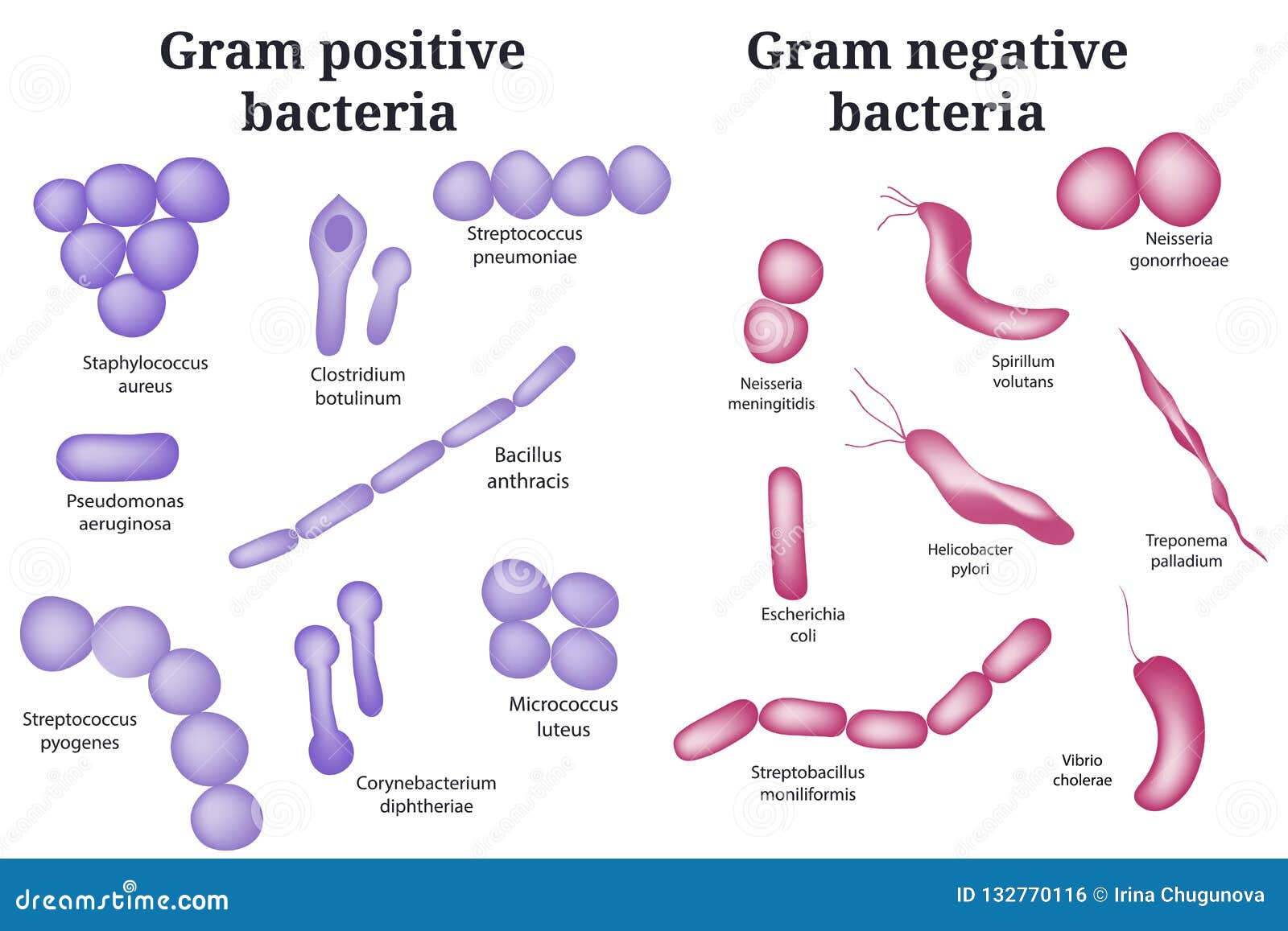
Gram positive bacteria vs gram negative bacteria (similarities and differences between gram positive and gram negative bacteria). They retain the color of crystal violet and stain dark blue or purple. Gram positive bacteria have a thick peptidoglycan layer and no outer lipid membrane whilst gram negative bacteria have a thin peptidoglycan layer gram positive bacteria have a distinctive purple appearance when observed under a light microscope following gram staining. It accounts 50% or more of the dry weight of the wall of some gram positive bacteria. The following article provides you the differentiation between them on the basis of various characteristics. Compared to gram positive bacteria that lack the external membrane, the peptidoglycan layer in gram negative bacteria is surrounded by an outer membrane. Lipid and lipoprotein content is high in the cell wall of gram negative bacteria. Few pathogenic bacteria belong to gram positive group. High murein content in cell wall. When comparing with the gram positive bacteria, gram negative bacteria are. Gram negative bacteria do not have this layer and thus do not retain the stain. Some of the differences are: Around 20 to 25 nm.
It accounts 50% or more of the dry weight of the wall of some gram positive bacteria. The classification is based on the bacterium's chemical these are bacteria that are not able to retain the crystal violet color and show negative result to gram stain test. Teichoic acid is present in many bacteria. Retain color of primary satin(crystal violet dye) and appears purple or blue. When comparing with the gram positive bacteria, gram negative bacteria are.
gram negative and gram positive bacteria - Google Search ... from i.pinimg.com Bacteria have cell walls made up of polysaccharides that give them strength and rigidity. Gram positive vs gram negative bacteria gram staining is a very important lab test. Few pathogenic bacteria belong to gram positive group. Gram positive bacteria retain the crystal violet stain during gram staining, giving the positive result. Teichoic acid is present in many bacteria. Thicker than gram negative bacteria. It is a faster approach compared to. They retain the color of crystal violet and stain dark blue or purple.
Generally thinner, 11 to 15 nm.
Bacteria of the genus haemophilus and gram negative bacteria stain pink when subjected to a gram stain procedure. They do not form a dispute, and in many cases are conditionally pathogenic. Gram positive bacteria have a thick peptidoglycan layer and no outer lipid membrane whilst gram negative bacteria have a thin peptidoglycan layer gram positive bacteria have a distinctive purple appearance when observed under a light microscope following gram staining. Bacteria are microscopic organisms, typically a few micrometers in length. Gram positive bacteria vs gram negative bacteria (similarities and differences between gram positive and gram negative bacteria). Gram positive vs gram negative bacteria gram staining is a very important lab test. The classification is based on the bacterium's chemical these are bacteria that are not able to retain the crystal violet color and show negative result to gram stain test. It paves way to the differentiation of the two distinct bacterial species. For this reason, the peptidoglycan layer is not the outermost part of the cell wall in these organisms. Gram staining technique is the most along with their staining characteristics, gram positive and gram negative bacteria differ from each other in various aspects which are listed below It is a faster approach compared to. Some of the differences are: Few pathogenic bacteria belong to gram positive group.
Lipid and lipoprotein content is high in the cell wall of gram negative bacteria. The gram method allowed to divide all microorganisms into two large groups. When comparing with the gram positive bacteria, gram negative bacteria are. Gram staining technique is the most along with their staining characteristics, gram positive and gram negative bacteria differ from each other in various aspects which are listed below The following article provides you the differentiation between them on the basis of various characteristics.
Gram Positive And Gram Negative Bacteria. Stock Vector ... from thumbs.dreamstime.com While it's similar to the plasma membrane. Few pathogenic bacteria belong to gram positive group. Diaminopamelic acid of one glycan backbone. Bacteria have cell walls made up of polysaccharides that give them strength and rigidity. This is due to retention. Bacteria are microscopic organisms, typically a few micrometers in length. The following article provides you the differentiation between them on the basis of various characteristics. Lipid and lipoprotein content is high in the cell wall of gram negative bacteria.
Gram negative coccobacillus bacteria have bacterial shapes that are in between spherical and rod shaped.
This is important since bacteria often experience variations in. High murein content in cell wall. Their ability to resist traditional antibiotics make them more dangerous. The outer membrane is made of lipopolysaccharide and staining or colour difference: What are the structural differences between gram positive and gram negative bacteria? While it's similar to the plasma membrane. Toxin production it is more accurate to write under gram negative bacteria exotoxins and/or endotoxins rather than exotoxins or endotoxins because endotoxins are produced by. With exception of neisseria, the rest. They are characterized by their cell envelopes, which are composed of a thin peptidoglycan cell wall sandwiched between an inner cytoplasmic cell. Bacteria of the genus haemophilus and gram negative bacteria stain pink when subjected to a gram stain procedure. Gram positive bacteria retain the crystal violet stain during gram staining, giving the positive result. They are decolorized when washed with absolute alcohol and acetone during gram staining. Gram positive bacteria have a layer of peptidoglycan in their cell wall that is notable for its ability to retain a grain stain (a complex formed between crystal violent and iodine).
You have just read the article entitled Gram Positive And Gram Negative Bacteria : Schematic views of Gram negative (top) and Gram positive ... / Hence, appear in purple or hence, gram negative bacteria appear in the secondary stain colour which is pink.. You can also bookmark this page with the URL : https://hag-io.blogspot.com/2021/05/gram-positive-and-gram-negative.html
Share Awesome
Belum ada Komentar untuk "Gram Positive And Gram Negative Bacteria : Schematic views of Gram negative (top) and Gram positive ... / Hence, appear in purple or hence, gram negative bacteria appear in the secondary stain colour which is pink."

Belum ada Komentar untuk "Gram Positive And Gram Negative Bacteria : Schematic views of Gram negative (top) and Gram positive ... / Hence, appear in purple or hence, gram negative bacteria appear in the secondary stain colour which is pink."
Posting Komentar